Guidelines for aging bovine pregnancies.
Keywords: hand, measurement, abortion, pregnancy, diagnosis, bovine, placentomes, crown-rump
It is not the author's intention to foist a personal bias onto colleagues; rather, to provide another system for students to entertain. Over the years, those who work with cattle develop methods that work for each of us; differing from one operator to the next. The methods mentioned here arise from the author's experience.
In this entry, the terms "pregnancy" and "gestation" are used interchangeably where appropriate.
In the absence of ultrasonography, an excellent way of improving aging accuracy is to ask a farmer not to provide the breeding date before an attempt is made at aging the pregnancy. It should be possible to determine the age of a pregnancy within at least, half of the duration of an estrous cycle (~10.5 days). In this manner, the farmer can determine the "dry-off" date without losing production or shortening the dry period unnecessarily. Between 42 and perhaps 90 days of gestation, it is frequently possible to determine the duration of gestation within four or five days but as the duration of gestation lengthens, one's accuracy decreases. Fortunately, the first third of gestation is usually the most critical for dairy farmers; not only must pregnancy be confirmed with certainty but in cases of re-breeding, it is often necessary to determine which insemination has accounted for the pregnancy.
There is arguably, only one cardinal sign of pregnancy i.e. fetal membrane slip. Enlargement of the uterus can be due many things other than pregnancy. Fremitus of the uterine artery can persist after calving or abortion and placentomes are palpable in those cases as well. Therefore, only after a fetal membrane slip has been detected, should one proceed to estimate the age of a pregnancy.
It is incorrect to refer to a "pregnant" horn because the fetal membranes on the side of ovulation occupy the contralateral horn by 18 to 20 days of gestation. Nevertheless, this convention persists and for convenience, is also used in this LORI entry.
In the image below (its detail only becoming evident when enlarged) the author's hand is shown as though it has grasped the pregnant horn in each case. The diameter of the uterus is represented by the area between the thumb and forefinger. Measurements for average male hands (the author's hands) and average female hands are shown at lower right in the image. Readers are encouraged to become familiar with the size of their own hands.
It is important to realize that a heifer's uterus is perhaps ten to twenty percent smaller than that of a multiparous cow at the same stage of pregnancy. Appropriate accommodation must be made for this fact when estimating the duration of gestation in a heifer. The growing uteruses in this image approximate those of a cow, not a heifer.

Image size: 2849x 2020px
The author has found it to be of great value to acquaint himself with the measurements of his hands, not only for estimating the duration of pregnancy or the age of aborted fetuses but for many other estimations too. Once one becomes acquainted with the dimensions of one's hands, their accuracy as measuring instruments are remarkable.
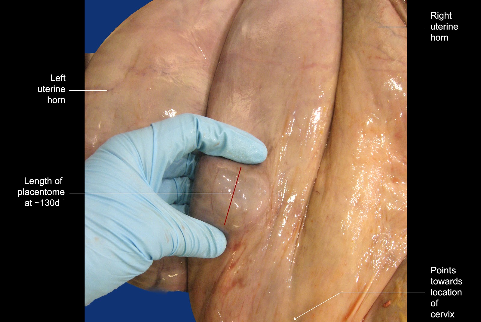
The image below shows the general area in which placentomes are palpated. In essence, one only uses the placentomes close to the cervix to make estimations of the size of these structures. If one palpates more cranial than this, it is likely that the duration of the pregnancy will be over estimated because placentomes generally become larger as one's hand moves cranially over the “pregnant” horn.

Image size: 3660x 2112px
Although the third image shows a placentome conveniently placed between thumb and forefinger for a measurement, this is not always possible in practice. More often, to gain an impression of their average size, several placentomes are manipulated between one's fingers while pressing down within the rectum.
The image below shows how placentomes grow during pregnancy. Note that their growth is almost logarithmic in the last third of gestation. The benchmarks discussed in the paragraph below are colored green in the image.

Image size: 1492x956px
At 75 days, placentomes usually become discernible for the first time.
At 90 days (3 months) placentomes measure about 1 x 1.5 cm in size. In the author's rather unconventional mind, it is easily remembered because 1.5 is half of 3 but admittedly, this is barely rational.
At 120 days (4 months) placentomes measure approximately 2.5 x 1.5 cm in size. Added together, 2.5 and 1.5 equal 4 ; easy to memorize.
At 150 days (5 months) placentomes measure approximately 2 x 3 cm in size. Added together, 2 and 3 equal 5 ; easy to memorize.
After 5 months, the remarkable growth rate of placentomes become noticeable.
The dimensions of placentomes at 5 months of gestation are particularly useful to memorize because they are about that size in beef cattle during routine fall pregnancy diagnosis. Simply by spreading one's fingers slightly and applying pressure on the dorsal surface of the uterus, then “raking’ in a cranial to caudal motion, a five month old pregnancy can be diagnosed easily. This procedure is so rapid and simple that hundreds beef cows can be examined in a few hours
At 6 months of gestation, the placentomes are approximately 6 cm long; again this is easy to memorize.
At term, the rapid growth of placentomes has increased their average length to approximately13 cm. Again, in the author's unconventional mind, this is remembered as “unlucky 13 at term".
Again, these benchmarks are 75 days, 3 months, 4 months, 5 months, 6 months and Term. Other durations are extrapolated between these benchmarks.
_____________________________
In the event that a fetus is aborted and one has to determine at what stage of gestation that occurred, many different guidelines are provided in the literature, some bordering on the ludicrous (small mouse, large rat without a tail etc). Instead, the following simple guidelines are offered. Again, one uses the dimensions of one's hand to make these determinations. A discussion follows the image.

Image size: 3046x2437px