Sexing the bovine fetus.
Keywords: sexing, bovine, ultrasound, male, female, ultrasonography
Image size: 640 x 480px
A female bovine features seen via transrectal ultrasonography at approximately 60 days of gestation. It is commonly states that the genital tubercle is used to determine gender in such cases. In this case for example, a caption could read: "The two highly echogenic spots visible between the hind legs, close to the tail show the location of the genital tubercle (arrow) in a female fetus. In the case of the male, the genital tubercle is seen as a bright echogenic structure just caudal to the abdominal attachment of the umbilicus."
However, the genital tubercle is not a paired structure, it develops into the clitoris in females and the penis in males. By contrast, the urogenital folds are paired structures (in both sexes) located on either side of the genital tubercle. Therefore it more likely that the paired echo seen above is being generated by the urogenital folds, not the genital tubercle.
The genital tubercle can be seen below. It is the precursor of the clitoris and is a single, sickle-shaped structure The urogenital ridges have almost developed into the vulva lips in this 60 day old bovine fetus (below).

Image size: 1263 x 927 px
The mammary gland (under circle) of this 60 day old fetus.
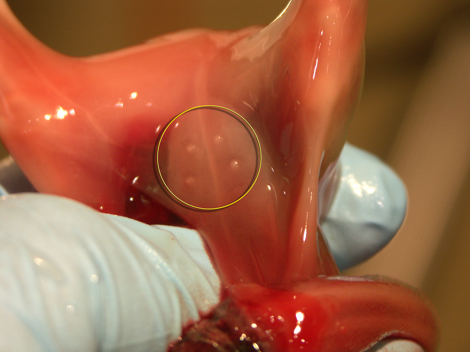
Image size: 1500 x 1125px
Although rudimentary teats are present in males, the mammary gland is well developed in females and serves as a landmark for sexing female fetuses. It should be visible via ultrasonography between 75 and 120 days of gestation.